
Effect of APOSEC on chronic ischemic left ventricular dysfunction
Long-acting beneficial effect of percutaneously intramyocardially delivered secretome of apoptotic peripheral blood cells on porcine chronic ischemic left ventricular dysfunction
Biomaterials. 2014 Jan 16. pii: S0142-9612(13)01567-6. doi: 10.1016/j.biomaterials.2013.12.071. [Epub ahead of print]
Pavo N, Zimmermann M, Pils D, Mildner M, Petrási Z, Petneházy Ö, Fuzik J, Jakab A, Gabriel C, Sipos W, Maurer G, Gyöngyösi M, Ankersmit HJ
Abstract
Background. The quantity of cells with paracrine effects for use in myocardial regeneration therapy is limited. Treatment with APOSEC may be more feasible and efficacious. This study investigated the effects of catheter-based endomyocardial injection of APOSEC (secretome of 2.5×10⁹ apoptotic peripheral blood mononuclear cells) on porcine chronic post-myocardial infarction (MI) left ventricular (LV) dysfunction and on gene expression.
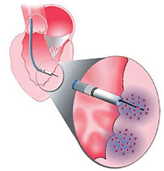
Methods. Closed-chest reperfused MI was induced in pigs by 90-min occlusion followed by reperfusion of the mid-LAD (day 0). At day 30, animals were randomized to receive porcine APOSEC (n=8) or medium solution (control; n=8) injected intramyocardially into the MI border zone using 3D NOGA guidance. At day 60, cardiac MRI with late enhancement and diagnostic NOGA (myocardial viability) was performed.Gene expression profiling of the infarct core, border zone, and normal myocardium was performed using microarray analysis and confirmed by quantitative real-time PCR.
Results. Injection of APOSEC significantly decreased infarct size (p<0.05) and improved cardiac index and myocardial viability compared to control. A trend towards higher LV ejection fraction was observed in APOSEC vs. control (45.4±5.9% vs. 37.4±8.9%, p=0.052). Transcriptome analysis revealed significant downregulation of caspase-1, tumor necrosis factor and other inflammatory genes in APOSEC affected areas. PCR showed higher expression of myogenic factor Mefc2 (p<0.05) and downregulated caspase-3 genes (p<0.05) in APOSEC-treated pigs.
Conclusions. Overexpression of MEF2c and repression of caspase-3 was related to decreased infarct size and improved cardiac function in secretome-treated animals. Altered gene expression 1-month post-APOSEC treatment suggested the long-acting effects of injected paracrine factors.
Keywords: cell-free regeneration therapy, genes, myocardial infarction, ischemia, remodeling
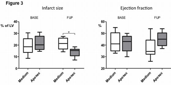
Part of Figure 3 is shown here. Ejection fraction (EF) at day 3 (BASE) and at day 60 (follow-up; FUP) showing a significant increase in EF at FUP in the APOSEC-treated pigs (n=8) compared to the control pigs (n=8). The APOSEC-treated pigs showed significantly smaller infarct size and better cardiac indexes. There was significantly lower end-diastolic pressure and end-diastolic stiffness in the APOSEC-treated group. *p<0.05